HL Paper 3
This question is about the use of ultrasound for diagnostic imaging.
Outline how ultrasound is produced for use in diagnostic imaging.
In order to look for damage to the chambers of the heart, ultrasound is used to form an image of the heart.
Suggest why it is better to use ultrasound rather than X-rays.
The speed of sound in skin is about five times the speed of sound in air. Given that the density of skin is about 700 times that of the density of air, compare the acoustic impedance of skin to that of air.
Explain, using your answer to (c), why, in using ultrasound for imaging, a layer of gel is placed between the transducer and the skin.
A wide range of frequencies of ultrasound may be used to image internal body organs. The choice of frequency for imaging a particular organ is determined by the depth of the organ beneath the skin.
Outline, with reference to attenuation and resolution, why the depth of the organ determines the choice of ultrasound frequency.
This question is about medical imaging.
A patient is suspected of having a partial blockage in his intestine as it leads away from his stomach. Possible medical imaging techniques include X-ray photography, ultrasound and the use of an endoscope.
When producing the X-ray photograph, the dose is kept to a minimum by a technique called enhancement.
(i) Outline why the dose needs to be kept to a minimum.
(ii) Describe one possible enhancement technique.
(iii) Discuss any extra procedures that are needed to get an appropriate image of the intestine in this situation.
A successful ultrasound scan relies on changes of acoustic impedance around the structure being imaged.
(i) Define acoustic impedance.
(ii) State the SI unit in which it is measured.
(iii) Explain, in terms of acoustic impedance, why gel needs to be applied on the surface of the skin before the ultrasound scan.
This question is about X-rays.
Define half-value thickness.
The half-value thickness in tissue for X-rays of a specific energy is 3.50 mm. Determine the fraction of the incident intensity of X-rays that has been transmitted through tissue of thickness 6.00 mm.
For X-rays of higher energy than those in (b), the half-value thickness is greater than 3.50 mm. State and explain the effect, if any, of this change on your answer in (b).
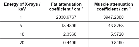
The attenuation values for fat and muscle at different X-ray energies are shown.
Outline the formation of a B scan in medical ultrasound imaging.
State what is meant by half-value thickness in X-ray imaging.
A monochromatic X-ray beam of energy 20 keV and intensity I0 penetrates 5.00 cm of fat and then 4.00 cm of muscle.
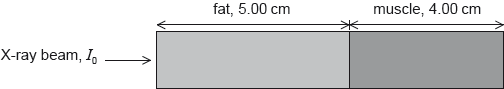
Calculate, in terms of I0, the final beam intensity that emerges from the muscle.
Compare the use of high and low energy X-rays for medical imaging.
State one advantage and one disadvantage of magnetic resonance imaging (MRI) compared to X-ray imaging.
Explain why a gradient field is required in nuclear magnetic resonance (NMR) imaging.
State the property of protons used in nuclear magnetic resonance (NMR) imaging.
Explain how a gradient field and resonance are produced in NMR to allow for the formation of images at a specific plane.
The density of muscle is 1075 kg m–3 and the speed of ultrasound in muscle is 1590 m s–1.
State a typical frequency used in medical ultrasound imaging.
Describe how an ultrasound transducer produces ultrasound.
Calculate the acoustic impedance Z of muscle.
Ultrasound of intensity 0.012 W\(\,\)cm–2 is incident on a water–muscle boundary. The acoustic impedance of water is 1.50 x 10⁶ kg\(\,\)m–2\(\,\)s–1.
The fraction of the incident intensity that is reflected is given by
\(\frac{{{{\left( {{Z_2} - {Z_1}} \right)}^2}}}{{{{\left( {{Z_2} + {Z_1}} \right)}^2}}}\)
where Z1 and Z2 are the acoustic impedances of medium 1 and medium 2.
Calculate the intensity of the reflected signal.
Some optic fibres consist of a core surrounded by cladding as shown in the diagram.

Calculate the maximum angle β for light to travel through the fibre.
Refractive index of core = 1.50
Refractive index of cladding = 1.48
Outline how the combination of core and cladding reduces the overall dispersion in the optic fibres.
The table shows the speed of ultrasound and the acoustic impedance for different media.
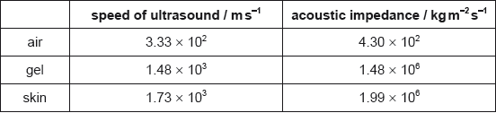
The fraction F of the intensity of an ultrasound wave reflected at the boundary between two media having acoustic impedances Z1 and Z2 is given by F = \(\frac{{{{({{\text{Z}}_1} - {{\text{Z}}_2})}^2}}}{{{{({{\text{Z}}_1} + {{\text{Z}}_2})}^2}}}\).
Outline how ultrasound is generated for medical imaging.
Describe one advantage and one disadvantage of using high frequencies ultrasound over low frequencies ultra sound for medical imaging.

Suggest one reason why doctors use ultrasound rather than X-rays to monitor the development of a fetus.
Calculate the density of skin.
Explain, with appropriate calculations, why a gel is used between the transducer and the skin.
This question is about nuclear magnetic resonance (NMR).
In nuclear magnetic resonance imaging, the patient is placed in a large uniform magnetic field. In addition, the part of the patient under investigation is subject to a weaker non-uniform (gradient) field.
Explain the role of these two fields in the imaging process.
In nuclear magnetic resonance (NMR) imaging radio frequency electromagnetic radiation is detected by the imaging sensors. Discuss the origin of this radiation.
This question is about transmission of digital signals in an optic fibre.
The input power to a single optic fibre X is 25 mW. The signal needs to be amplified when the power has been attenuated to 4.0 ×10⁻¹⁹ W. The attenuation loss in the optic fibre is 1.8 dB km–1.
Calculate the maximum distance between amplifiers in the system.
This question is about X-rays.
(i) X-rays travelling in a medium experience attenuation. State what is meant by attenuation.
(ii) Show that the half-value thickness \({x_{\frac{1}{2}}}\) is related to the attenuation coefficient \(\mu \) by
\[\mu {x_{\frac{1}{2}}} = 1{\rm{n}}2\]
(iii) Estimate the fraction of the incident intensity of an X-ray beam that has travelled through 2.0 cm of muscle. The half-value thickness of muscle is 0.73 cm.
This question is about nuclear magnetic resonance (NMR) imaging.
Outline the physical principles of NMR imaging.
State two advantages of NMR imaging over computed tomography (CT) imaging.
1.
2.
This question is about the use of ultrasound.
Define acoustic impedance.
State the significance of acoustic impedance in the use of ultrasound techniques.
Medical practitioners select the frequency of the ultrasound depending on the diagnosis they are undertaking. Outline the importance of using ultrasound of the appropriate frequency.
This question is about ultrasound.
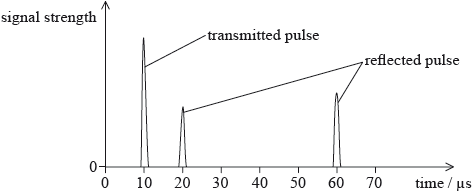
An ultrasound pulse is transmitted into the body of a patient. The pulse is partially reflected at a fat–muscle boundary and then, deeper in the body, at a muscle–bone boundary. The graph shows the variation with time of the signal strength at the transducer.
Muscle has density \(1.08 \times {10^3}{\text{ kg}}\,{{\text{m}}^{ - 3}}\) and acoustic impedance \(1.70 \times {10^6}{\text{ kg}}\,{{\text{m}}^{ - 2}}{{\text{s}}^{ - 1}}\).
Define acoustic impedance
(i) Calculate the speed of ultrasound in muscle.
(ii) Determine the thickness of the muscle layer in the patient.
State one advantage and one disadvantage of using ultrasound of frequency 1 MHz, rather than 3 MHz, in medical diagnosis.
Advantage:
Disadvantage:
This question is about medical imaging using X-rays.
The table shows the attenuation coefficient \(\mu \) (mu) for different parts of the body.
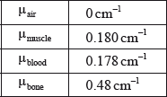
An X-ray scan is taken of a patient to examine the flow of blood through their arm. X-rays of intensity \(I\) are incident on an equal thickness of blood and muscle. The intensity of the X-rays is measured after passing through the blood and muscle respectively.
Define attenuation coefficient.
Calculate the half-value thickness for blood.
Calculate the ratio \(\frac{{{I_{{\text{blood}}}}}}{{{I_{{\text{muscle}}}}}}\) for 1 cm of tissue.
Suggest why an X-ray scan does not allow for the differentiation between muscle and blood.
A contrast medium containing iodine is injected into the patient. This increases the attenuation coefficient of blood so that the difference between the intensities of blood and muscle is greater than 2%. The blood can now be observed on an X-ray scan. Determine the minimum increase in \({\mu _{{\text{blood}}}}\) that will enable a sharper contrast to be observed between an equal thickness of muscle and blood.
X-rays are a form of ionizing radiation. To reduce the danger to a patient, the intensity of X-rays are kept to a minimum. Describe how enhancement allows for low intensity X-rays to be used.
An X-ray beam of intensity I0 is incident on lead. After travelling a distance x through the lead the intensity of the beam is reduced to I.
The graph shows the variation of ln\(\left( {\frac{I}{{{I_0}}}} \right)\) with x.

Show that the attenuation coefficient of lead is 60 cm–1.
A technician operates an X-ray machine that takes 100 images each day. Estimate the width of the lead screen that is required so that the total exposure of the technician in 250 working days is equal to the exposure that the technician would receive from one X-ray exposure without the lead screen.
The diagram represents a simple optical astronomical reflecting telescope with the path of some light rays shown.

It is proposed to build an array of radio telescopes such that the maximum distance between them is 3800 km. The array will operate at a wavelength of 2.1 cm.
Comment on whether it is possible to build an optical telescope operating at 580 nm that is to have the same resolution as the array.
This question is about X-rays.
Two parallel beams of monochromatic X-rays of the same intensity are incident on equal thicknesses of bone and fat in a patient.

The attenuation coefficient for fat is 180m−1 and the attenuation coefficient for bone is 345m−1. The thickness of both materials is 0.0150m.
Calculate \(\frac{{{I_b}}}{{{I_f}}}\) where \({I_b}\) is the intensity of the beam leaving the bone and \({I_f}\) is the intensity of the beam leaving the fat.
Explain how fluorescent emitters are used to enhance the image formed on a photographic X-ray plate.
This question is about X-rays.
Define the attenuation coefficient as applied to a beam of X-rays travelling through a medium.
Derive the relationship between the attenuation coefficient μ and the half-value thickness \({x_{\frac{1}{2}}}\).
Aluminium is often used to filter out the low energy X-rays in a beam of X-rays. The following data are available for a particular X-ray beam.

Assuming equal initial intensities, determine, after the X-ray beam has passed through an aluminium sheet 6.0 mm thick, the following ratio.
\[\frac{{{\rm{intensity of 15keV X - rays}}}}{{{\rm{intensity of 30keV X - rays}}}}\]
Outline why X-rays are not suitable to image an organ such as the liver.
In the context of nuclear magnetic resonance (NMR) imaging explain the role of
Outline why the fracture in a broken bone can be seen in a medical X-ray image.
The diagram shows X-rays incident on tissue and bone.

The thicknesses of bone and tissue are both 0.054 m.
The intensity of X-rays transmitted through bone is Ib and the intensity transmitted through tissue is It.
The following data are available.
Mass absorption coefficient for bone = mass absorption
coefficient for tissue = 1.2 × 10⁻²\(\,\)m2\(\,\)kg–1
Density of bone = 1.9 × 10³ kg\(\,\)m–3
Density of tissue = 1.1 × 10³ kg\(\,\)m–3
Calculate the ratio \(\frac{{{I_{\text{b}}}}}{{{I_{\text{t}}}}}\).
the large uniform magnetic field applied to the patient.
the radio-frequency signal emitted towards the patient.
the non-uniform magnetic field applied to the patient.
This question is about medical imaging.
The intensity of a parallel X-ray beam is reduced to 50% of its initial intensity when it passes through bone of thickness 1.2 cm. Determine the thickness of bone needed to reduce the intensity of the same X-ray beam to 15% of its initial value.
The linear attenuation coefficient μ of a material is affected by the energy of the X-ray beam and by the density ρ of the material. The mass absorption coefficient is equal to \(\frac{\mu }{\rho }\) to take into account the density of the material.
The graph shows the variation of mass absorption coefficient with energy of the X-ray beam for both muscle and bone.

Show that the attenuation coefficient for bone of density 1800 kg m–3, for X-rays of 20 keV, is about 7 cm–1.
The density of muscle is 1200 kg m–3. Calculate the ratio of intensities to compare, for a beam of 20 keV, the attenuation produced by 1 cm of bone and 1 cm of muscle.
Suggest why more energetic beams of about 150 keV would be unsuitable for imaging a bone–muscle section of a body.
This question is about ultrasound scanning.
Outline how ultrasound is generated for medical diagnostic purposes.
When ultrasound of intensity I0 travels in a medium of acoustic impedance Z1 and is incident on a medium of acoustic impedance Z2, the intensity IR that is reflected at the interface is given by the following equation.
\[{I_R} = {\left( {\frac{{{Z_1} - {Z_2}}}{{{Z_1} + {Z_2}}}} \right)^2}{I_0}\]
The following data are available.

Use the data to deduce why a layer of gel must be used between a transducer and the patient’s skin in medical ultrasound imaging.
In medical scanning, practitioners have the option of using A-scans or B-scans. Distinguish, with reference to the techniques used to produce the scans, between an A-scan and a B-scan.
This question is about X-rays.
Define attenuation coefficient.
The graph shows how the attenuation coefficient μ of muscle varies with photon energy E.

In X-ray imaging, photons of energy less than 20 ke V are filtered out of the beam.
(i) Explain, with reference to the graph, why this does not significantly affect the quality of the X-ray image produced.
(ii) State the advantage to the patient of filtering out the low energy photons from the X-ray beam.
(iii) Calculate the fraction of the intensity transmitted through 3.0 mm of muscle for X-rays of energy 50 ke V.
This question is about X-ray absorption.
A parallel beam of X-rays is incident on a section of tissue of thickness x. The constant incident intensity is I0 and the transmitted intensity is It.

The half-value thickness of the tissue is 4.0 cm.
On the axes below, sketch a graph to show the variation with tissue thickness x of \(\frac{{{I_t}}}{{{I_0}}}\).

Calculate the attenuation coefficient of X-rays for this tissue.
For a different type of tissue, the ratio \(\frac{{{I_t}}}{{{I_0}}}\) is smaller for the same thickness x of material.
Compare the attenuation coefficient of this tissue with that of the tissue in (b).
Barium has an attenuation coefficient that is much larger than that for human tissue.
Explain why a patient is asked to drink a liquid barium meal to help produce an X-ray image of the digestive system.
This question is about X-rays.
Define attenuation coefficient.
The graph below shows the variation of attenuation coefficient μ with photon energy E for X-rays in an absorbing medium.

A beam of X-rays is incident on a sample of the medium with intensity I0. Using the graph,
(i) determine how far X-rays with energy equal to 0.1MeV travel inside the sample before their intensity reduces to 0.1I0.
(ii) predict whether X-rays of energy 10MeV are more penetrating than X-rays of energy 0.1MeV in this medium.
This question is about ultrasound.
Define acoustic impedance of a medium.
The acoustic impedances for various media are shown in the table.

Ultrasound is incident normally on a layer of soft tissue. Gel is placed between the skin and the transducer.

The fraction of the intensity of ultrasound that is reflected (reflection coefficient) at the boundary of two media of impedances Z1 and Z2 is given by the following equation.
\[{\left( {\frac{{{Z_2} - {Z_1}}}{{{Z_2} + {Z_1}}}} \right)^2}\]
(i) Suggest why the gel allows the ultrasound to enter the soft tissue without any reflection.
(ii) Calculate the reflection coefficient at the soft tissue–bone boundary.
(iii) The soft tissue between the skin and the bone absorbs 60% of the intensity of ultrasound travelling through it. The intensity of ultrasound leaving the transducer is I0. Determine, in terms of I0, the intensity of the ultrasound that is reflected back into the transducer from the bone.
This question is about ultrasound.
The diagram shows part of a cross-section through the leg of a patient who is undergoing an ultrasound scan.

Data for the speed c of ultrasound in different media are shown below, together with values for the acoustic impedance Z.

Use the data from the table to calculate a value for the density of bone.
The fraction F of the intensity of an ultrasound wave reflected at the boundary between two media having acoustic impedances
Z1 and Z2 is given by the following equation.
\[F = \frac{{{{\left( {{Z_1} - {Z_2}} \right)}^2}}}{{{{\left( {{Z_1} + {Z_2}} \right)}^2}}}\]
Determine the fraction F for the boundary between
(i) air and muscle.
(ii) gel and muscle.
Use your answers in (b) to explain the need for a gel on the patient’s skin.
This question is about the use of X-rays and ultrasound in medical imaging.
The diagram below shows X-rays being used to scan a sample of bone and muscle.

(i) Outline how the arrangement differentiates between bone and muscle.
(ii) Use the data below to determine the ratio \(\frac{{{I_b}}}{{{{\mathop{\rm I}\nolimits} _m}}}\) where Ib and Im are the intensity of X-rays reaching the photographic plate through the bone and the muscle, respectively.
Thickness x of sample = 10.0 cm
Linear attenuation coefficient of bone μb = 0.53 cm–1
Linear attenuation coefficient of muscle μm = 0.30 cm–1
(iii) The half-value thickness of a material increases as the energy of the radiation increases.
Discuss, with reference to penetration and effect on tissue, why using low energy X-rays in medical imaging is highly desirable but is rare in practice.
The same sample is now investigated with an ultrasound A-scan from the side as shown.

(i) State one advantage of ultrasound over X-ray imaging.
(ii) State why gel is needed at the transducer-muscle boundary.
(iii) A short pulse is directed from the transducer into the sample at time t = 0.
The graph shows how the intensity of the reflected signal from the muscle-bone boundary varies as a function of time. The speed of sound in muscle is 1.6×10³ m s–1.

Calculate the thickness y of the sample of muscle.
This question is about ultrasonic imaging.
The table gives the velocity of sound in, and the densities of, the materials.

(i) State the SI unit for acoustic impedance.
(ii) Calculate the acoustic impedance for each material and write your answers in the table above.
In medical imaging, X-rays can be passed through aluminium before reaching the body. The graph shows the variation of the linear absorption coefficient of aluminium for different photon energies.

X-rays are incident on an aluminium sheet of thickness 8.0 cm. Calculate the fraction of the incident X-ray intensity that emerges from this sheet for photon energies of
(i) 9.0 MeV.
(ii) 3.0×10⁻³ MeV.
With reference to your answers to (a)(i) and (a)(ii), discuss the advantages of using the aluminium sheet.
This question is about medical imaging.
Define attenuation coefficient.
X-rays are used in dentistry to reveal decay inside teeth. In a research study, a tooth is partially filled with a new glass-based material to replace decayed tissue. X-ray photographs are taken of the tooth.

A parallel beam of X-rays is incident on the tooth. X-rays emerging at A have travelled through enamel and filling only, X-rays emerging at B have travelled through enamel and tooth tissue only.
(i) Show that the ratio \(\frac{{{\rm{intensity of X - rays at A}}}}{{{\rm{intensity of X - rays at B}}}}\) is approximately 3×10⁻⁷.
(ii) The X-ray exposure time is such as to enable fine detail in the enamel to be revealed by X-rays emerging at B. Suggest, with reference to the ratio in (b)(i), why the contrast at B is much greater than the contrast at A.
A complete dental record of all the teeth in a patient’s mouth requires about 20 separate X-ray exposures. Image intensifiers are now used in dentistry to allow a single image to be made of all the teeth with one exposure. Outline the advantages of this for the patient.
The table shows data about the acoustic impedance of some materials that would be involved in the transmission of ultrasound through a tooth.

Without carrying out a calculation, outline two reasons why ultrasound is not used to detect the presence of decay inside a tooth.